
Heart Anatomical Model B Ultrasound Color Doppler Ultrasound Cardiology Organs
$59.19 NZD
Approx $34.51 USD
Description:
Introduction to the Heart Anatomical Model with B-Mode and Color Doppler Ultrasound Visualization
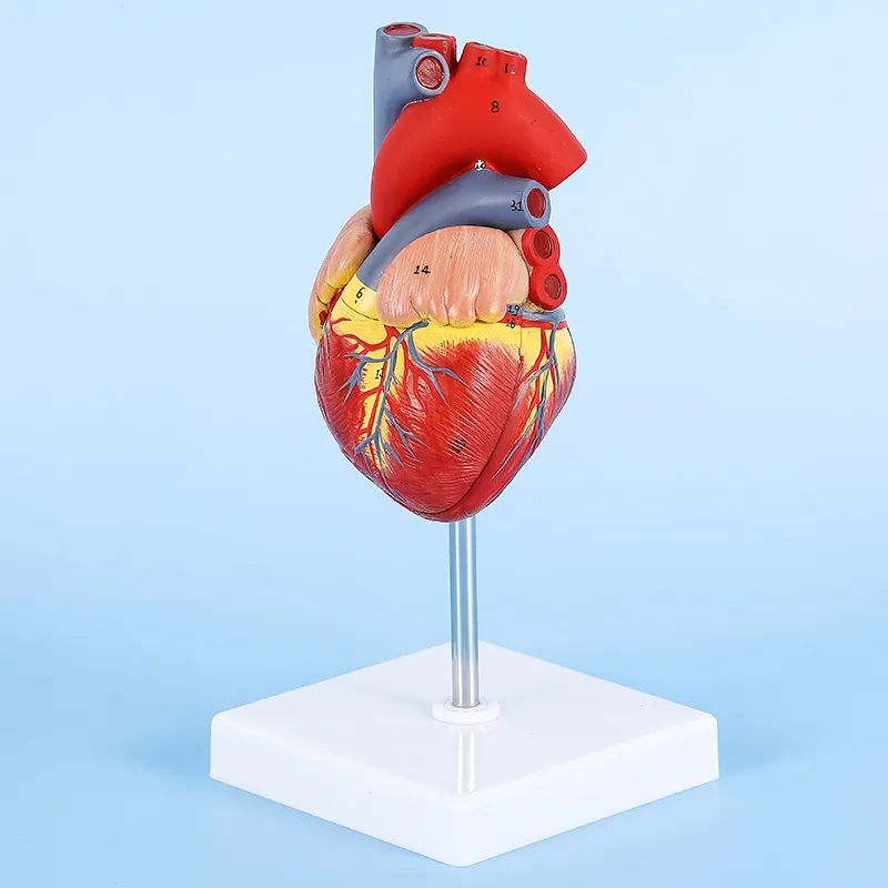
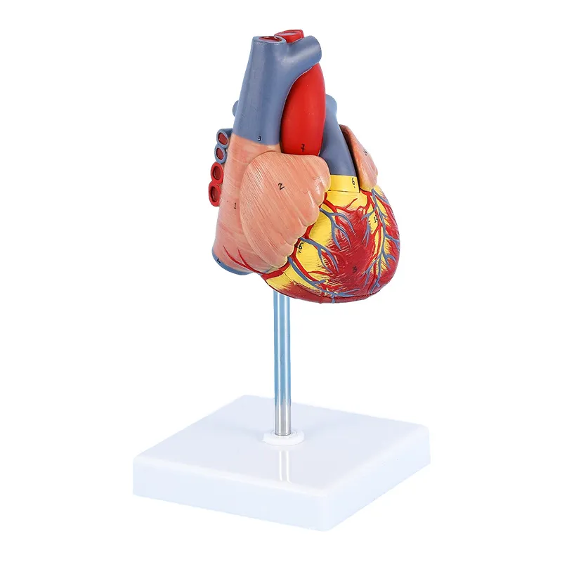
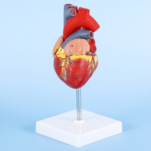
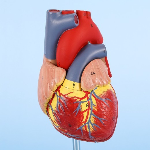
The Heart Anatomical Model with B-Mode and Color Doppler Ultrasound Visualization is a unique educational tool designed to bridge heart anatomy and ultrasound imaging. This model includes a detailed heart replica with illustrated Doppler ultrasound imaging overlays, making it ideal for understanding both structural anatomy and echocardiographic visualization. Perfect for medical students, cardiologists, and educators, this model aids in the study of heart anatomy, cardiac function, and the interpretation of Doppler ultrasound imaging. From classrooms to cardiology clinics across New Zealand, this model supports hands-on learning in cardiac health and ultrasound diagnostics.
H2: Key Features of the Heart Anatomical Model with Ultrasound Visualization
1. Realistic Heart Anatomy with High Detail
This model accurately represents the anatomy of the human heart, including the atria, ventricles, valves, coronary arteries, and other essential structures. Each feature is crafted to reflect its shape, location, and function, allowing students and professionals to study the heart’s structure and understand how blood flows through its chambers.
2. Illustrated B-Mode and Color Doppler Ultrasound Views
The model includes illustrated overlays of B-mode (grayscale) and Color Doppler ultrasound images, showing how heart structures appear in diagnostic imaging. B-mode imaging highlights structural details, while Color Doppler imaging shows blood flow dynamics, with color-coded visuals representing flow direction and velocity. This feature helps users learn to interpret ultrasound images in a real-world clinical context, making it an ideal tool for echocardiography training.
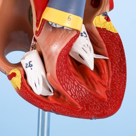
3. Detailed Visualization of Heart Valves and Blood Flow
The Color Doppler illustrations allow users to see how blood flows through the heart, depicting areas where turbulence or blockage might occur. The model highlights key features such as the mitral, aortic, tricuspid, and pulmonary valves, showing their role in maintaining unidirectional blood flow. This detailed visualization is invaluable for understanding heart function and diagnosing valve-related conditions.
4. Color-Coded and Labeled Sections for Easy Identification
Each anatomical structure and ultrasound overlay is color-coded and labeled to help users identify and differentiate heart structures and understand blood flow patterns. This color-coding enhances comprehension, making it easier to identify each part of the heart and the respective ultrasound markers. This feature is especially useful for visual learners and those new to cardiology or echocardiography.
5. High-Quality, Durable Construction
Made from non-toxic, high-grade materials, this heart model is built to withstand regular handling in both educational and clinical settings. The model’s sturdy construction ensures that it maintains its structural integrity and detail over time, making it suitable for anatomy classrooms, ultrasound labs, and cardiology clinics in New Zealand. The durability ensures a long-lasting educational resource for heart anatomy and ultrasound training.
6. Compact and Display-Ready Design
The model is compact and can be displayed on desks, lab tables, or shelves, making it accessible for individual study and group demonstrations. Its stable base keeps it secure, allowing for hands-on interaction in both clinical and classroom environments. This portability and display-ready design make it an excellent tool for educators and healthcare providers alike.
H2: Why Choose the Heart Anatomical Model with Ultrasound Visualization?
1. Essential for Cardiology and Echocardiography Education
This model is an indispensable tool for teaching cardiology and echocardiography, especially for students and professionals studying the structural and functional aspects of the heart. By combining physical heart anatomy with ultrasound overlays, it provides a well-rounded understanding of cardiac function and diagnostic imaging, helping students in New Zealand master foundational knowledge in cardiology.
2. Ideal for Ultrasound and Diagnostic Imaging Training
In ultrasound and diagnostic imaging programs, this model is invaluable for understanding heart structures and interpreting Color Doppler and B-mode imaging. Trainers can use it to explain how ultrasound imaging visualizes the heart, blood flow, and common conditions like valve regurgitation or stenosis, making it ideal for those focused on cardiac ultrasound diagnostics.
3. Supports Visual and Kinesthetic Learning
This model caters to both visual and kinesthetic learners, offering a hands-on experience that enhances comprehension. Color-coded sections support visual learning by clearly defining heart structures and ultrasound markers, while the physical model allows kinesthetic learners to interact directly with heart anatomy, making complex information easier to grasp.
4. Useful for Patient Education in Clinical Settings
In clinical settings, this model is ideal for educating patients about heart anatomy, blood flow, and ultrasound imaging. By visually explaining conditions and diagnostic imaging techniques, healthcare providers can help patients understand their heart health, the significance of imaging results, and potential treatment options. This model supports effective communication and informed decision-making.
5. Educational Display for Clinics and Classrooms
Beyond its educational function, the Heart Anatomical Model with Ultrasound Visualization serves as an informative display for classrooms, labs, and cardiology clinics. In these environments, it provides a visual aid for discussions about cardiac health, blood flow, and diagnostic imaging, making it ideal for educational settings that focus on cardiology and diagnostics.
H2: Maintenance and Care Tips for Your Heart Anatomical Model with Ultrasound Visualization
To keep your model in excellent condition, follow these care tips:
-
Dust Regularly: Use a soft cloth or brush to remove dust from the model, especially around detailed areas like valves and
ultrasound overlays. Regular cleaning helps maintain the model’s appearance and prevents dirt buildup.
-
Avoid Direct Sunlight: Display the model out of direct sunlight to prevent colors from fading over time, preserving the
vibrancy of both the anatomical and ultrasound illustrations.
-
Handle with Care: Although durable, handle the model gently to avoid wear or damage, particularly around delicate parts
like the valves and ultrasound illustrations.
- Store Properly When Not in Use: When not displayed, store the model in its original packaging or a dust-free area to protect it from potential damage. Proper storage helps extend the model’s lifespan for long-term educational use.
Product information:
Material: PVC
Product name: 1 to 1 heart model
Product material: PVC
Use scene: indoor
Use crowd: adult
Size Information:
Size: 11*11*22.5 cm
Packing list:
Decoration x1